How to use
- Decide which model to use:
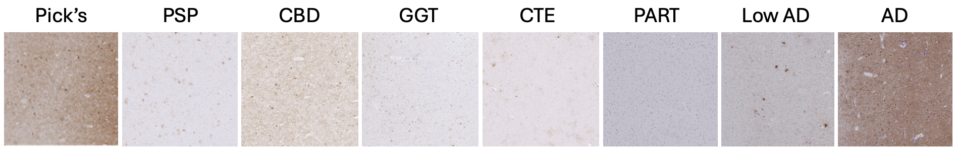
Low magnification (5×) = overall disease pattern
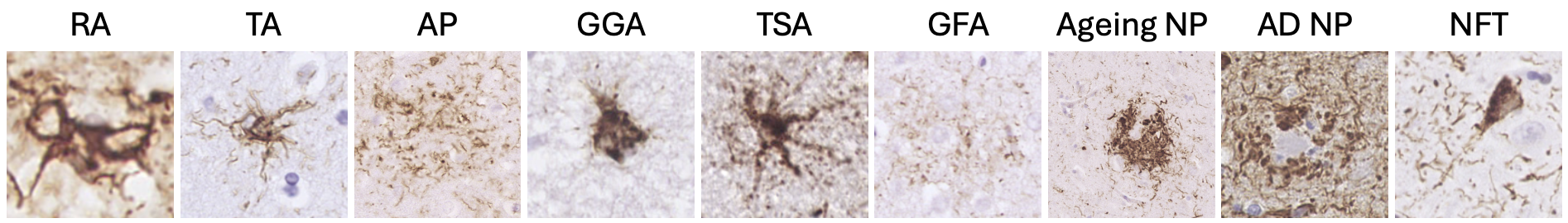
High magnification (40×) = individual aggregates
- Take snapshots:
Extract square snapshots from the region of interest at the correct magnification.
- Prepare your images:
Save snapshots into a single folder.
Ensure images match the selected model:
• Low magnification – representative field of the region
• High magnification – one aggregate per image
- Upload images:
Select the folder to upload. Multiple images will be processed automatically.
- Interpret outputs:
The model returns class probabilities (%) for each image.
Higher values indicate stronger morphological similarity to a trained class.
- Export results:
Download predictions as an Excel file for further analysis.
*Models were trained on AT8-stained tau pathology images, but may generalise to other tau immunostains.
*Predictions are probabilistic outputs from a supervised convolutional neural network and should be interpreted alongside full neuropathological assessment, not used as a standalone diagnostic tool.